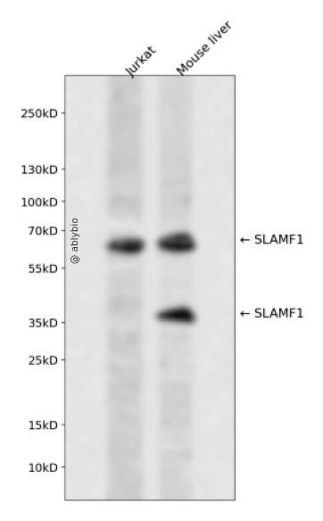
Western blot analysis of SLAMF1 expressed in Jurkat,Mouse liver using SLAMF1 Rabbit pAb at 1:1000. Secondary antibody: HRP Goat Anti-Rabbit IgG (H+L) at 1:5000. Lysates/proteins: 30ug per lane. Blocking buffer: 5% non-fat dry milk in TBST. Detection: ECL Enhanced Kit. Exposure time: 120s.
Western blot analysis of SLAMF1 expressed in Jurkat,Mouse liver using SLAMF1 Rabbit pAb at 1:1000. Secondary antibody: HRP Goat Anti-Rabbit IgG (H+L) at 1:5000. Lysates/proteins: 30ug per lane. Blocking buffer: 5% non-fat dry milk in TBST. Detection: ECL Enhanced Kit. Exposure time: 120s.
SLAM-induced signal-transduction events in T-lymphocytes are different from those in B-cells. Two modes ofSLAM signaling are likely to exist: one in which the inhibitor SH2D1A acts as a negative regulator and another inwhich protein-tyrosine phosphatase 2C (PTPN11)-dependent signal transduction operates.
基因ID
6504
基因名
SLAMF1
Swiss
Q13291
别名
SLAMF1;CD150;CDw150;SLAM
组织表达
Constitutively expressed on peripheral blood memory T-cells, T-cell clones, immature thymocytes and a proportion of B-cells, and is rapidly induced on naive T-cells after activation (PubMed:7617038). Activated B-cells express isoform 1, isoform 3 and a cytoplasmic isoform (PubMed:9091591). Isoform 4 is expressed in B-cells, primary T-cells, dendritic cells and macrophages. Isoform 4 is expressed in tumors of the central nervous system (PubMed:25710480).
功能
Self-ligand receptor of the signaling lymphocytic activation molecule (SLAM) family. SLAM receptors triggered by homo- or heterotypic cell-cell interactions are modulating the activation and differentiation of a wide variety of immune cells and thus are involved in the regulation and interconnection of both innate and adaptive immune response. Activities are controlled by presence or absence of small cytoplasmic adapter proteins, SH2D1A/SAP and/or SH2D1B/EAT-2. SLAMF1-induced signal-transduction events in T-lymphocytes are different from those in B-cells. Two modes of SLAMF1 signaling seem to exist: one depending on SH2D1A (and perhaps SH2D1B) and another in which protein-tyrosine phosphatase 2C (PTPN11)-dependent signal transduction operates. Initially it has been proposed that association with SH2D1A prevents binding to inhibitory effectors including INPP5D/SHIP1 and PTPN11/SHP-2 (PubMed:11806999). However, signaling is also regulated by SH2D1A which can simultaneously interact with and recruit FYN which subsequently phosphorylates and activates SLAMF1 (PubMed:12458214). Mediates IL-2-independent proliferation of activated T-cells during immune responses and induces IFN-gamma production (By similarity). Downstreaming signaling involves INPP5D, DOK1 and DOK2 leading to inhibited IFN-gamma production in T-cells, and PRKCQ, BCL10 and NFKB1 leading to increased T-cell activation and Th2 cytokine production (By similarity). Promotes T-cell receptor-induced IL-4 secretion by CD4+ cells (By similarity). Inhibits antigen receptor-mediated production of IFN-gamma, but not IL-2, in CD4-/CD8- T-cells (By similarity). Required for IL-4 production by germinal centers T follicular helper (T(Fh))cells (By similarity). May inhibit CD40-induced signal transduction in monocyte-derived dendritic cells (PubMed:16317102). May play a role in allergic responses and may regulate allergen-induced Th2 cytokine and Th1 cytokine secretion (By similarity). In conjunction with SLAMF6 controls the transition between positive selection and the subsequent expansion and differentiation of the thymocytic natural killer T (NKT) cell lineage. Involved in the peripheral differentiation of indifferent natural killer T (iNKT) cells toward a regulatory NKT2 type (By similarity). In macrophages involved in down-regulation of IL-12, TNF-alpha and nitric oxide in response to lipopolysaccharide (LPS) (By similarity). In B-cells activates the ERK signaling pathway independently of SH2D1A but implicating both, SYK and INPP5D, and activates Akt signaling dependent on SYK and SH2D1A (By similarity). In B-cells also activates p38 MAPK and JNK1 and JNK2 (PubMed:20231852). In conjunction with CD84/SLAMF5 and SLAMF6 may be a negative regulator of the humoral immune response (By similarity). Involved in innate immune response against Gram-negative bacteria in macrophages; probably recognizes OmpC and/or OmpF on the bacterial surface, regulates phagosome maturation and recruitment of the PI3K complex II (PI3KC3-C2) leading to accumulation of PdtIns3P and NOX2 activity in the phagosomes (PubMed:20818396).
a. 贴壁培养细胞收集
去除贴壁细胞的培养液,用PBS、NS或无血清培养基清洗1次,低速离心,弃上清,留取沉淀。
b. 悬浮培养细胞收集
速离心悬浮细胞,弃上清,收集沉淀。手指轻弹细胞,使其松散。
c. 组织样本收集
把组织剪切成细小的碎片,越小越好。取液氮或超低温冰箱中冷冻30min以上的组织,迅速用液氮研磨,研磨过程尽量控制在1~2min之内,以减少蛋白的降解。
(2)总蛋白提取
a. 细胞/组织裂解
将装有细胞沉淀或组织碎片的容器完全插入冰中。细胞沉淀按照1mL裂解液/107个细胞(1个T75培养瓶细胞量)的比例加入相应体积的裂解液(细胞量足够时都加入3mL,不足时根据细胞量计算),裂解20min,每隔5min将离心管置于涡旋振荡仪上震荡10s。组织碎片按照0.5mL 裂解液/100mg组织向匀浆器中加入蛋白裂解液,每3min研磨一次,重复5次,使组织尽量碾碎。(裂解液中根据需要选择添加或不添加蛋白酶抑制剂)。
b. 离心
把裂解好的样品配平后,置于预冷的高速冷冻离心机中,12000 rpm,15min。
c. 蛋白变性
完成离心后,上清即为蛋白提取液。吸取少量蛋白提取液做蛋白浓度测定。向剩余的蛋白提取液的离心管中加入1/5上清体积的5×Loading Buffer(最终工作液为1X),待干式恒温器温度升至95℃后,将1.5mL离心管插入加热孔中,95℃加热变性10min,待液体完全冷却后置于-20℃保存。
(3)蛋白浓度测定(BCA法)
a. BCA工作液的配置
根据样品数量,按50体积BCA试剂A加入1体积BCA试剂B(50:1)配置适量BCA工作液,充分混匀。BCA工作液室温24h内稳定。
b. 标准品测定
取10μl蛋白标准品(5mg/ml BSA)稀释至50μl,使终浓度为1mg/ml。稀释后的蛋白标准品可以-20℃长期保存。此标准品溶液的稀释液可使用去离子水或1*PBS。将标准品按0、1、2、4、8、12、16、20μl加入到96孔板中,加稀释液补足到20μl(见附表)。加适当体积样品到96孔板的样品孔中,如果样本不足20μl,需加稀释液补足到20μl。请注意记录样品体积。各孔加入200μl BCA工作液,37℃放置20-30min。用酶标仪测定A562,或540-595nm之间的其他波长吸光度。根据标准曲线和使用的样品体积计算出样品的蛋白浓度。
a. Western Transfer Buffer至少提前2h (即开始电泳后)放入-20℃冰箱预冷,但注意避免结冰。
b. 根据胶体大小,将Filter Paper及Nitrocellulose membrane剪裁至合适尺寸。
c. 目的蛋白>20KD选择0.45μm NC膜/PVDF膜;目的蛋白<20KD选择0.2μmNC膜或PVDF膜,选择完毕后将NC膜放在Western Transfer Buffer中浸泡备用,注意如使用的是PVDF膜需先放入甲醇中浸泡5-10min,再放入Western Transfer Buffer中浸泡备用。
(2)裂解液&洗杂液:Cell lysis buffer for IP (without inhibitors)
(3)蛋白酶抑制剂
(4)封闭液:含 3% BSA 的 1X PBS
(5)1×PBS 缓冲液
(6)5×loding buffer(使用时用去离子水稀释至工作浓度即可)
(7)Control IgG (AC005/ AC011/AC034)
二、实验步骤
1、样本处理
(1)贴壁培养细胞
a. 取裂解液室温溶解混匀,根据需要选择添加或不添加蛋白酶抑制剂。
b. 去除贴壁细胞的培养液,用PBS、NS或无血清培养基清洗1次,低速离心,弃上清,留取沉淀。
c. 按照6孔板每孔加入100~200μl裂解液的比例,加入裂解液。移液器轻轻吹打,使裂解液和细胞充分接触。通常裂解液作用于细胞1~5s内,细胞会被裂解。
d. 1000~12000g,离心3~5min(如果用冷冻离心机4℃效果更佳),取上清。
(2)悬浮培养细胞
a. 取裂解液室温溶解混匀,根据需要选择添加或不添加蛋白酶抑制剂。
b. 速离心悬浮细胞,弃上清,收集沉淀。
c. 手指轻弹细胞,使其松散。按照6孔板每孔加入100~200μl裂解液的比例,加入NP-40裂解液。通常6孔板每孔加入100~200μl裂解液已经足够,但如果细胞密度非常高可以适当加大裂解液的用量150~200μl,再用手指轻弹以充分裂解细胞。充分裂解后应无明显沉淀。
d. 1000~12000g,离心3~5min(如果用冷冻离心机4℃效果更佳),取上清。
(3)组织样本
a. 取裂解液室温溶解混匀,根据需要选择添加或不添加蛋白酶抑制剂。
b. 把组织剪切成细小的碎片,越小越好。
c. 取液氮或超低温冰箱中冷冻30min以上的组织,迅速用液氮研磨,研磨过程尽量控制在1~2min之内,以减少蛋白的降解。
d. 按照每20mg组织加入100~200μl裂解液的比例,加入含有PMSF的裂解液。冰上或4℃裂解30-60min。(步骤3、4也可采用以下过程:按照每20mg组织加入100~200μl裂解液的比例加入NP-40裂解液。用玻璃匀浆器或组织研磨器匀浆,直至充分裂解,过程尽量控制在1~2min之内,以减少蛋白的降解。)
e. 按照每20mg组织加入100~200μl裂解液的比例,加入裂解液。
f. 1000~12000g,4℃离心10~15min(如无低温离心机,室温下离心也可),取上清。
2、磁珠预处理
(1)将rProtein A/G Plus MaqPoly Beads颠倒或漩涡混匀,翻转瓶身发现底部无黑色沉淀即可。
(2)取30μl rProtein A/G Plus MaqPoly Beads至新的EP管中,放在磁分离器上,待溶液澄清后,用移液器吸弃保护液。
(3)将EP管从磁分离器上取下来,加入1ml Cell lysis buffer for IP (without inhibitors),混匀,放置在磁分离器上,收集磁珠,用移液器吸弃洗杂液,重复2次。