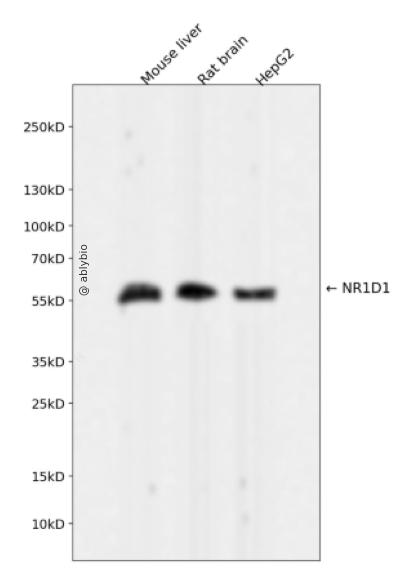
Western blot analysis of NR1D1 expressed in Mouse liver,Rat brain,HepG2 using NR1D1 Rabbit mAb at 1:1000. Secondary antibody: HRP Goat Anti-Rabbit IgG (H+L) at 1:5000. Lysates/proteins: 30ug per lane. Blocking buffer: 5% non-fat dry milk in TBST. Detection: ECL Enhanced Kit. Exposure time: 120s.
Western blot analysis of NR1D1 expressed in Mouse liver,Rat brain,HepG2 using NR1D1 Rabbit mAb at 1:1000. Secondary antibody: HRP Goat Anti-Rabbit IgG (H+L) at 1:5000. Lysates/proteins: 30ug per lane. Blocking buffer: 5% non-fat dry milk in TBST. Detection: ECL Enhanced Kit. Exposure time: 120s.
This gene encodes a transcription factor that is a member of the nuclear receptor subfamily 1. The encoded protein is a ligand-sensitive transcription factor that negatively regulates the expression of core clock proteins. In particular this protein represses the circadian clock transcription factor aryl hydrocarbon receptor nuclear translocator-like protein 1 (ARNTL). This protein may also be involved in regulating genes that function in metabolic, inflammatory and cardiovascular processes.
Widely expressed. Expressed at high levels in the liver, adipose tissue, skeletal muscle and brain. Also expressed in endothelial cells (ECs), vascular smooth muscle cells (VSMCs) and macrophages. Expression oscillates diurnally in the suprachiasmatic nucleus (SCN) of the hypothalamus as well as in peripheral tissues. Expression increases during the differentiation of pre-adipocytes into mature adipocytes. Expressed at high levels in some squamous carcinoma cell lines.
功能
Transcriptional repressor which coordinates circadian rhythm and metabolic pathways in a heme-dependent manner. Integral component of the complex transcription machinery that governs circadian rhythmicity and forms a critical negative limb of the circadian clock by directly repressing the expression of core clock components ARTNL/BMAL1, CLOCK and CRY1. Also regulates genes involved in metabolic functions, including lipid and bile acid metabolism, adipogenesis, gluconeogenesis and the macrophage inflammatory response. Acts as a receptor for heme which stimulates its interaction with the NCOR1/HDAC3 corepressor complex, enhancing transcriptional repression. Recognizes two classes of DNA response elements within the promoter of its target genes and can bind to DNA as either monomers or homodimers, depending on the nature of the response element. Binds as a monomer to a response element composed of the consensus half-site motif 5'-[A/G]GGTCA-3' preceded by an A/T-rich 5' sequence (RevRE), or as a homodimer to a direct repeat of the core motif spaced by two nucleotides (RevDR-2). Acts as a potent competitive repressor of ROR alpha (RORA) function and regulates the levels of its ligand heme by repressing the expression of PPARGC1A, a potent inducer of heme synthesis. Regulates lipid metabolism by repressing the expression of APOC3 and by influencing the activity of sterol response element binding proteins (SREBPs); represses INSIG2 which interferes with the proteolytic activation of SREBPs which in turn govern the rhythmic expression of enzymes with key functions in sterol and fatty acid synthesis. Regulates gluconeogenesis via repression of G6PC and PEPCK and adipocyte differentiation via repression of PPARG. Regulates glucagon release in pancreatic alpha-cells via the AMPK-NAMPT-SIRT1 pathway and the proliferation, glucose-induced insulin secretion and expression of key lipogenic genes in pancreatic-beta cells. Positively regulates bile acid synthesis by increasing hepatic expression of CYP7A1 via repression of NR0B2 and NFIL3 which are negative regulators of CYP7A1. Modulates skeletal muscle oxidative capacity by regulating mitochondrial biogenesis and autophagy; controls mitochondrial biogenesis and respiration by interfering with the STK11-PRKAA1/2-SIRT1-PPARGC1A signaling pathway. Represses the expression of SERPINE1/PAI1, an important modulator of cardiovascular disease and the expression of inflammatory cytokines and chemokines in macrophages. Represses gene expression at a distance in macrophages by inhibiting the transcription of enhancer-derived RNAs (eRNAs). Plays a role in the circadian regulation of body temperature and negatively regulates thermogenic transcriptional programs in brown adipose tissue (BAT); imposes a circadian oscillation in BAT activity, increasing body temperature when awake and depressing thermogenesis during sleep. In concert with NR2E3, regulates transcriptional networks critical for photoreceptor development and function. In addition to its activity as a repressor, can also act as a transcriptional activator. In the ovarian granulosa cells acts as a transcriptional activator of STAR which plays a role in steroid biosynthesis. In collaboration with SP1, activates GJA1 transcription in a heme-independent manner.
a. 贴壁培养细胞收集
去除贴壁细胞的培养液,用PBS、NS或无血清培养基清洗1次,低速离心,弃上清,留取沉淀。
b. 悬浮培养细胞收集
速离心悬浮细胞,弃上清,收集沉淀。手指轻弹细胞,使其松散。
c. 组织样本收集
把组织剪切成细小的碎片,越小越好。取液氮或超低温冰箱中冷冻30min以上的组织,迅速用液氮研磨,研磨过程尽量控制在1~2min之内,以减少蛋白的降解。
(2)总蛋白提取
a. 细胞/组织裂解
将装有细胞沉淀或组织碎片的容器完全插入冰中。细胞沉淀按照1mL裂解液/107个细胞(1个T75培养瓶细胞量)的比例加入相应体积的裂解液(细胞量足够时都加入3mL,不足时根据细胞量计算),裂解20min,每隔5min将离心管置于涡旋振荡仪上震荡10s。组织碎片按照0.5mL 裂解液/100mg组织向匀浆器中加入蛋白裂解液,每3min研磨一次,重复5次,使组织尽量碾碎。(裂解液中根据需要选择添加或不添加蛋白酶抑制剂)。
b. 离心
把裂解好的样品配平后,置于预冷的高速冷冻离心机中,12000 rpm,15min。
c. 蛋白变性
完成离心后,上清即为蛋白提取液。吸取少量蛋白提取液做蛋白浓度测定。向剩余的蛋白提取液的离心管中加入1/5上清体积的5×Loading Buffer(最终工作液为1X),待干式恒温器温度升至95℃后,将1.5mL离心管插入加热孔中,95℃加热变性10min,待液体完全冷却后置于-20℃保存。
(3)蛋白浓度测定(BCA法)
a. BCA工作液的配置
根据样品数量,按50体积BCA试剂A加入1体积BCA试剂B(50:1)配置适量BCA工作液,充分混匀。BCA工作液室温24h内稳定。
b. 标准品测定
取10μl蛋白标准品(5mg/ml BSA)稀释至50μl,使终浓度为1mg/ml。稀释后的蛋白标准品可以-20℃长期保存。此标准品溶液的稀释液可使用去离子水或1*PBS。将标准品按0、1、2、4、8、12、16、20μl加入到96孔板中,加稀释液补足到20μl(见附表)。加适当体积样品到96孔板的样品孔中,如果样本不足20μl,需加稀释液补足到20μl。请注意记录样品体积。各孔加入200μl BCA工作液,37℃放置20-30min。用酶标仪测定A562,或540-595nm之间的其他波长吸光度。根据标准曲线和使用的样品体积计算出样品的蛋白浓度。
a. Western Transfer Buffer至少提前2h (即开始电泳后)放入-20℃冰箱预冷,但注意避免结冰。
b. 根据胶体大小,将Filter Paper及Nitrocellulose membrane剪裁至合适尺寸。
c. 目的蛋白>20KD选择0.45μm NC膜/PVDF膜;目的蛋白<20KD选择0.2μmNC膜或PVDF膜,选择完毕后将NC膜放在Western Transfer Buffer中浸泡备用,注意如使用的是PVDF膜需先放入甲醇中浸泡5-10min,再放入Western Transfer Buffer中浸泡备用。
(2)裂解液&洗杂液:Cell lysis buffer for IP (without inhibitors)
(3)蛋白酶抑制剂
(4)封闭液:含 3% BSA 的 1X PBS
(5)1×PBS 缓冲液
(6)5×loding buffer(使用时用去离子水稀释至工作浓度即可)
(7)Control IgG (AC005/ AC011/AC034)
二、实验步骤
1、样本处理
(1)贴壁培养细胞
a. 取裂解液室温溶解混匀,根据需要选择添加或不添加蛋白酶抑制剂。
b. 去除贴壁细胞的培养液,用PBS、NS或无血清培养基清洗1次,低速离心,弃上清,留取沉淀。
c. 按照6孔板每孔加入100~200μl裂解液的比例,加入裂解液。移液器轻轻吹打,使裂解液和细胞充分接触。通常裂解液作用于细胞1~5s内,细胞会被裂解。
d. 1000~12000g,离心3~5min(如果用冷冻离心机4℃效果更佳),取上清。
(2)悬浮培养细胞
a. 取裂解液室温溶解混匀,根据需要选择添加或不添加蛋白酶抑制剂。
b. 速离心悬浮细胞,弃上清,收集沉淀。
c. 手指轻弹细胞,使其松散。按照6孔板每孔加入100~200μl裂解液的比例,加入NP-40裂解液。通常6孔板每孔加入100~200μl裂解液已经足够,但如果细胞密度非常高可以适当加大裂解液的用量150~200μl,再用手指轻弹以充分裂解细胞。充分裂解后应无明显沉淀。
d. 1000~12000g,离心3~5min(如果用冷冻离心机4℃效果更佳),取上清。
(3)组织样本
a. 取裂解液室温溶解混匀,根据需要选择添加或不添加蛋白酶抑制剂。
b. 把组织剪切成细小的碎片,越小越好。
c. 取液氮或超低温冰箱中冷冻30min以上的组织,迅速用液氮研磨,研磨过程尽量控制在1~2min之内,以减少蛋白的降解。
d. 按照每20mg组织加入100~200μl裂解液的比例,加入含有PMSF的裂解液。冰上或4℃裂解30-60min。(步骤3、4也可采用以下过程:按照每20mg组织加入100~200μl裂解液的比例加入NP-40裂解液。用玻璃匀浆器或组织研磨器匀浆,直至充分裂解,过程尽量控制在1~2min之内,以减少蛋白的降解。)
e. 按照每20mg组织加入100~200μl裂解液的比例,加入裂解液。
f. 1000~12000g,4℃离心10~15min(如无低温离心机,室温下离心也可),取上清。
2、磁珠预处理
(1)将rProtein A/G Plus MaqPoly Beads颠倒或漩涡混匀,翻转瓶身发现底部无黑色沉淀即可。
(2)取30μl rProtein A/G Plus MaqPoly Beads至新的EP管中,放在磁分离器上,待溶液澄清后,用移液器吸弃保护液。
(3)将EP管从磁分离器上取下来,加入1ml Cell lysis buffer for IP (without inhibitors),混匀,放置在磁分离器上,收集磁珠,用移液器吸弃洗杂液,重复2次。