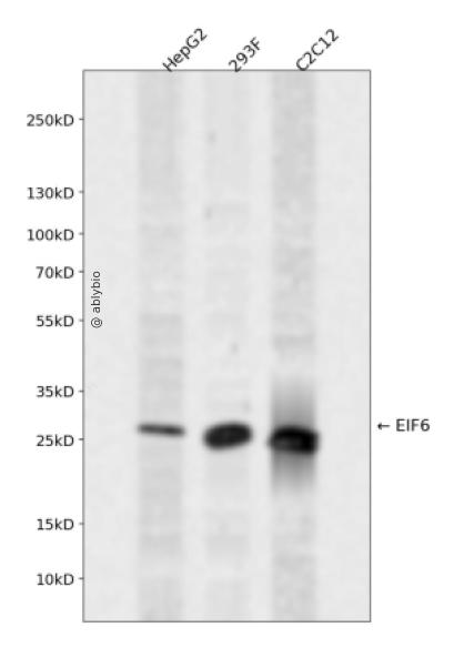
Western blot analysis of EIF6 expressed in HepG2,293F,C2C12 using EIF6 Rabbit pAb at 1:1000. Secondary antibody: HRP Goat Anti-Rabbit IgG (H+L) at 1:5000. Lysates/proteins: 30ug per lane. Blocking buffer: 5% non-fat dry milk in TBST. Detection: ECL Enhanced Kit. Exposure time: 120s.
Western blot analysis of EIF6 expressed in HepG2,293F,C2C12 using EIF6 Rabbit pAb at 1:1000. Secondary antibody: HRP Goat Anti-Rabbit IgG (H+L) at 1:5000. Lysates/proteins: 30ug per lane. Blocking buffer: 5% non-fat dry milk in TBST. Detection: ECL Enhanced Kit. Exposure time: 120s.
Hemidesmosomes are structures which link the basal lamina to the intermediate filament cytoskeleton. An important functional component of hemidesmosomes is the integrin beta-4 subunit (ITGB4), a protein containing two fibronectin type III domains. The protein encoded by this gene binds to the fibronectin type III domains of ITGB4 and may help link ITGB4 to the intermediate filament cytoskeleton. The encoded protein, which is insoluble and found both in the nucleus and in the cytoplasm, can function as a translation initiation factor and prevent the association of the 40S and 60S ribosomal subunits. Multiple non-protein coding transcript variants and variants encoding two different isoforms have been found for this gene.
Expressed at very high levels in colon carcinoma with lower levels in normal colon and ileum and lowest levels in kidney and muscle (at protein level).
功能
Binds to the 60S ribosomal subunit and prevents its association with the 40S ribosomal subunit to form the 80S initiation complex in the cytoplasm. Behaves as a stimulatory translation initiation factor downstream insulin/growth factors. Is also involved in ribosome biogenesis. Associates with pre-60S subunits in the nucleus and is involved in its nuclear export. Cytoplasmic release of TIF6 from 60S subunits and nuclear relocalization is promoted by a RACK1 (RACK1)-dependent protein kinase C activity (PubMed:10085284, PubMed:14654845, PubMed:21536732). In tissues responsive to insulin, controls fatty acid synthesis and glycolysis by exerting translational control of adipogenic transcription factors such as CEBPB, CEBPD and ATF4 that have G/C rich or uORF in their 5'UTR. Required for ROS-dependent megakaryocyte maturation and platelets formation, controls the expression of mitochondrial respiratory chain genes involved in reactive oxygen species (ROS) synthesis (By similarity). Involved in miRNA-mediated gene silencing by the RNA-induced silencing complex (RISC). Required for both miRNA-mediated translational repression and miRNA-mediated cleavage of complementary mRNAs by RISC (PubMed:17507929). Modulates cell cycle progression and global translation of pre-B cells, its activation seems to be rate-limiting in tumorigenesis and tumor growth (By similarity).
实验步骤
蛋白质印迹法标准操作流程
一、实验试剂及耗材
1、样品制备试剂
a. RIPA 裂解液
b. 蛋白酶抑制剂
c. BCA工作液
d. BSA Standard Solution(5mg/ml)
e. 1×PBS缓冲液
f. 5×loading buffer
2、制胶试剂
a.30% Acr-Bis (29:1)
b.1 M Tris-HCl (pH6.8)
C.1.5M Tris-HCl (pH8.8)
d.10%SDS
e.10%Ammonium persulfate (APS)
f.TEMED
3、电泳、转膜试剂及耗材
a. 1X Tris-Glycine SDS Running Buffer
b. 1X Western Transfer Buffer
c. Filter Paper (7.5×10cm)
d. Nitrocellulose membrane
4、封闭、一抗二抗孵育及曝光试剂
a. 1XTBST Buffer
b. 1X Western Blocking Buffer
c. Skimmed milk powder
d. Bovine Serum Albumin (BSA)
e. Western Antibody Dilution Buffer
f. HRP 偶联二抗(AS014/AS003)
g. SignalFire™ ECL Reagent
二、实验步骤
1、样品制备
(1)细胞收集
a. 贴壁培养细胞收集
去除贴壁细胞的培养液,用PBS、NS或无血清培养基清洗1次,低速离心,弃上清,留取沉淀。
b. 悬浮培养细胞收集
速离心悬浮细胞,弃上清,收集沉淀。手指轻弹细胞,使其松散。
c. 组织样本收集
把组织剪切成细小的碎片,越小越好。取液氮或超低温冰箱中冷冻30min以上的组织,迅速用液氮研磨,研磨过程尽量控制在1~2min之内,以减少蛋白的降解。
(2)总蛋白提取
a. 细胞/组织裂解
将装有细胞沉淀或组织碎片的容器完全插入冰中。细胞沉淀按照1mL裂解液/107个细胞(1个T75培养瓶细胞量)的比例加入相应体积的裂解液(细胞量足够时都加入3mL,不足时根据细胞量计算),裂解20min,每隔5min将离心管置于涡旋振荡仪上震荡10s。组织碎片按照0.5mL 裂解液/100mg组织向匀浆器中加入蛋白裂解液,每3min研磨一次,重复5次,使组织尽量碾碎。(裂解液中根据需要选择添加或不添加蛋白酶抑制剂)。
b. 离心
把裂解好的样品配平后,置于预冷的高速冷冻离心机中,12000 rpm,15min。
c. 蛋白变性
完成离心后,上清即为蛋白提取液。吸取少量蛋白提取液做蛋白浓度测定。向剩余的蛋白提取液的离心管中加入1/5上清体积的5×Loading Buffer(最终工作液为1X),待干式恒温器温度升至95℃后,将1.5mL离心管插入加热孔中,95℃加热变性10min,待液体完全冷却后置于-20℃保存。
(3)蛋白浓度测定(BCA法)
a. BCA工作液的配置
根据样品数量,按50体积BCA试剂A加入1体积BCA试剂B(50:1)配置适量BCA工作液,充分混匀。BCA工作液室温24h内稳定。
b. 标准品测定
取10μl蛋白标准品(5mg/ml BSA)稀释至50μl,使终浓度为1mg/ml。稀释后的蛋白标准品可以-20℃长期保存。此标准品溶液的稀释液可使用去离子水或1*PBS。将标准品按0、1、2、4、8、12、16、20μl加入到96孔板中,加稀释液补足到20μl(见附表)。加适当体积样品到96孔板的样品孔中,如果样本不足20μl,需加稀释液补足到20μl。请注意记录样品体积。各孔加入200μl BCA工作液,37℃放置20-30min。用酶标仪测定A562,或540-595nm之间的其他波长吸光度。根据标准曲线和使用的样品体积计算出样品的蛋白浓度。
a. Western Transfer Buffer至少提前2h (即开始电泳后)放入-20℃冰箱预冷,但注意避免结冰。
b. 根据胶体大小,将Filter Paper及Nitrocellulose membrane剪裁至合适尺寸。
c. 目的蛋白>20KD选择0.45μm NC膜/PVDF膜;目的蛋白<20KD选择0.2μmNC膜或PVDF膜,选择完毕后将NC膜放在Western Transfer Buffer中浸泡备用,注意如使用的是PVDF膜需先放入甲醇中浸泡5-10min,再放入Western Transfer Buffer中浸泡备用。
(2)裂解液&洗杂液:Cell lysis buffer for IP (without inhibitors)
(3)蛋白酶抑制剂
(4)封闭液:含 3% BSA 的 1X PBS
(5)1×PBS 缓冲液
(6)5×loding buffer(使用时用去离子水稀释至工作浓度即可)
(7)Control IgG (AC005/ AC011/AC034)
二、实验步骤
1、样本处理
(1)贴壁培养细胞
a. 取裂解液室温溶解混匀,根据需要选择添加或不添加蛋白酶抑制剂。
b. 去除贴壁细胞的培养液,用PBS、NS或无血清培养基清洗1次,低速离心,弃上清,留取沉淀。
c. 按照6孔板每孔加入100~200μl裂解液的比例,加入裂解液。移液器轻轻吹打,使裂解液和细胞充分接触。通常裂解液作用于细胞1~5s内,细胞会被裂解。
d. 1000~12000g,离心3~5min(如果用冷冻离心机4℃效果更佳),取上清。
(2)悬浮培养细胞
a. 取裂解液室温溶解混匀,根据需要选择添加或不添加蛋白酶抑制剂。
b. 速离心悬浮细胞,弃上清,收集沉淀。
c. 手指轻弹细胞,使其松散。按照6孔板每孔加入100~200μl裂解液的比例,加入NP-40裂解液。通常6孔板每孔加入100~200μl裂解液已经足够,但如果细胞密度非常高可以适当加大裂解液的用量150~200μl,再用手指轻弹以充分裂解细胞。充分裂解后应无明显沉淀。
d. 1000~12000g,离心3~5min(如果用冷冻离心机4℃效果更佳),取上清。
(3)组织样本
a. 取裂解液室温溶解混匀,根据需要选择添加或不添加蛋白酶抑制剂。
b. 把组织剪切成细小的碎片,越小越好。
c. 取液氮或超低温冰箱中冷冻30min以上的组织,迅速用液氮研磨,研磨过程尽量控制在1~2min之内,以减少蛋白的降解。
d. 按照每20mg组织加入100~200μl裂解液的比例,加入含有PMSF的裂解液。冰上或4℃裂解30-60min。(步骤3、4也可采用以下过程:按照每20mg组织加入100~200μl裂解液的比例加入NP-40裂解液。用玻璃匀浆器或组织研磨器匀浆,直至充分裂解,过程尽量控制在1~2min之内,以减少蛋白的降解。)
e. 按照每20mg组织加入100~200μl裂解液的比例,加入裂解液。
f. 1000~12000g,4℃离心10~15min(如无低温离心机,室温下离心也可),取上清。
2、磁珠预处理
(1)将rProtein A/G Plus MaqPoly Beads颠倒或漩涡混匀,翻转瓶身发现底部无黑色沉淀即可。
(2)取30μl rProtein A/G Plus MaqPoly Beads至新的EP管中,放在磁分离器上,待溶液澄清后,用移液器吸弃保护液。
(3)将EP管从磁分离器上取下来,加入1ml Cell lysis buffer for IP (without inhibitors),混匀,放置在磁分离器上,收集磁珠,用移液器吸弃洗杂液,重复2次。