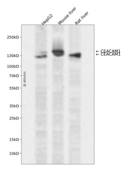
Western blot analysis of CEACAM1 expressed in HepG2,Mouse liver,Rat liver using CEACAM1 Rabbit pAb at 1:1000. Secondary antibody: HRP Goat Anti-Rabbit IgG (H+L) at 1:5000. Lysates/proteins: 30ug per lane. Blocking buffer: 5% non-fat dry milk in TBST. Detection: ECL Enhanced Kit. Exposure time: 120s.
Western blot analysis of CEACAM1 expressed in HepG2,Mouse liver,Rat liver using CEACAM1 Rabbit pAb at 1:1000. Secondary antibody: HRP Goat Anti-Rabbit IgG (H+L) at 1:5000. Lysates/proteins: 30ug per lane. Blocking buffer: 5% non-fat dry milk in TBST. Detection: ECL Enhanced Kit. Exposure time: 120s.
This gene encodes a member of the carcinoembryonic antigen (CEA) gene family, which belongs to the immunoglobulin superfamily. Two subgroups of the CEA family, the CEA cell adhesion molecules and the pregnancy-specific glycoproteins, are located within a 1.2 Mb cluster on the long arm of chromosome 19. Eleven pseudogenes of the CEA cell adhesion molecule subgroup are also found in the cluster. The encoded protein was originally described in bile ducts of liver as biliary glycoprotein. Subsequently, it was found to be a cell-cell adhesion molecule detected on leukocytes, epithelia, and endothelia. The encoded protein mediates cell adhesion via homophilic as well as heterophilic binding to other proteins of the subgroup. Multiple cellular activities have been attributed to the encoded protein, including roles in the differentiation and arrangement of tissue three-dimensional structure, angiogenesis, apoptosis, tumor suppression, metastasis, and the modulation of innate and adaptive immune responses. Multiple transcript variants encoding different isoforms have been reported, but the full-length nature of all variants has not been defined.
基因ID
634
基因名
CEACAM1
Swiss
P13688
别名
CEACAM1;BGP;BGP1;BGPI
组织表达
Expressed in columnar epithelial cells of the colon (at protein level) (PubMed:10436421). The predominant forms expressed by T cells are those containing a long cytoplasmic domain (PubMed:18424730). Expressed in granulocytes and lymphocytes. Leukocytes only express isoforms 6 and isoform 1 (PubMed:11994468).
功能
Isoform 1: Cell adhesion protein that mediates homophilic cell adhesion in a calcium-independent manner (By similarity). Plays a role as coinhibitory receptor in immune response, insulin action and functions also as an activator during angiogenesis (PubMed:18424730, PubMed:23696226, PubMed:25363763). Its coinhibitory receptor function is phosphorylation- and PTPN6 -dependent, which in turn, suppress signal transduction of associated receptors by dephosphorylation of their downstream effectors. Plays a role in immune response, of T cells, natural killer (NK) and neutrophils (PubMed:18424730, PubMed:23696226). Upon TCR/CD3 complex stimulation, inhibits TCR-mediated cytotoxicity by blocking granule exocytosis by mediating homophilic binding to adjacent cells, allowing interaction with and phosphorylation by LCK and interaction with the TCR/CD3 complex which recruits PTPN6 resulting in dephosphorylation of CD247 and ZAP70 (PubMed:18424730). Also inhibits T cell proliferation and cytokine production through inhibition of JNK cascade and plays a crucial role in regulating autoimmunity and anti-tumor immunity by inhibiting T cell through its interaction with HAVCR2 (PubMed:25363763). Upon natural killer (NK) cells activation, inhibit KLRK1-mediated cytolysis of CEACAM1-bearing tumor cells by trans-homophilic interactions with CEACAM1 on the target cell and lead to cis-interaction between CEACAM1 and KLRK1, allowing PTPN6 recruitment and then VAV1 dephosphorylation (PubMed:23696226). Upon neutrophils activation negatively regulates IL1B production by recruiting PTPN6 to a SYK-TLR4-CEACAM1 complex, that dephosphorylates SYK, reducing the production of reactive oxygen species (ROS) and lysosome disruption, which in turn, reduces the activity of the inflammasome. Downregulates neutrophil production by acting as a coinhibitory receptor for CSF3R by downregulating the CSF3R-STAT3 pathway through recruitment of PTPN6 that dephosphorylates CSF3R (By similarity). Also regulates insulin action by promoting INS clearance and regulating lipogenesis in liver through regulating insulin signaling (By similarity). Upon INS stimulation, undergoes phosphorylation by INSR leading to INS clearance by increasing receptor-mediated insulin endocytosis. This inernalization promotes interaction with FASN leading to receptor-mediated insulin degradation and to reduction of FASN activity leading to negative regulation of fatty acid synthesis. INSR-mediated phosphorylation also provokes a down-regulation of cell proliferation through SHC1 interaction resulting in decrease coupling of SHC1 to the MAPK3/ERK1-MAPK1/ERK2 and phosphatidylinositol 3-kinase pathways (By similarity). Functions as activator in angiogenesis by promoting blood vessel remodeling through endothelial cell differentiation and migration and in arteriogenesis by increasing the number of collateral arteries and collateral vessel calibers after ischemia. Also regulates vascular permeability through the VEGFR2 signaling pathway resulting in control of nitric oxide production (By similarity). Downregulates cell growth in response to EGF through its interaction with SHC1 that mediates interaction with EGFR resulting in decrease coupling of SHC1 to the MAPK3/ERK1-MAPK1/ERK2 pathway (By similarity). Negatively regulates platelet aggregation by decreasing platelet adhesion on type I collagen through the GPVI-FcRgamma complex (By similarity). Inhibits cell migration and cell scattering through interaction with FLNA; interfers with the interaction of FLNA with RALA (PubMed:16291724). Mediates bile acid transport activity in a phosphorylation dependent manner (By similarity). Negatively regulates osteoclastogenesis (By similarity).
a. 贴壁培养细胞收集
去除贴壁细胞的培养液,用PBS、NS或无血清培养基清洗1次,低速离心,弃上清,留取沉淀。
b. 悬浮培养细胞收集
速离心悬浮细胞,弃上清,收集沉淀。手指轻弹细胞,使其松散。
c. 组织样本收集
把组织剪切成细小的碎片,越小越好。取液氮或超低温冰箱中冷冻30min以上的组织,迅速用液氮研磨,研磨过程尽量控制在1~2min之内,以减少蛋白的降解。
(2)总蛋白提取
a. 细胞/组织裂解
将装有细胞沉淀或组织碎片的容器完全插入冰中。细胞沉淀按照1mL裂解液/107个细胞(1个T75培养瓶细胞量)的比例加入相应体积的裂解液(细胞量足够时都加入3mL,不足时根据细胞量计算),裂解20min,每隔5min将离心管置于涡旋振荡仪上震荡10s。组织碎片按照0.5mL 裂解液/100mg组织向匀浆器中加入蛋白裂解液,每3min研磨一次,重复5次,使组织尽量碾碎。(裂解液中根据需要选择添加或不添加蛋白酶抑制剂)。
b. 离心
把裂解好的样品配平后,置于预冷的高速冷冻离心机中,12000 rpm,15min。
c. 蛋白变性
完成离心后,上清即为蛋白提取液。吸取少量蛋白提取液做蛋白浓度测定。向剩余的蛋白提取液的离心管中加入1/5上清体积的5×Loading Buffer(最终工作液为1X),待干式恒温器温度升至95℃后,将1.5mL离心管插入加热孔中,95℃加热变性10min,待液体完全冷却后置于-20℃保存。
(3)蛋白浓度测定(BCA法)
a. BCA工作液的配置
根据样品数量,按50体积BCA试剂A加入1体积BCA试剂B(50:1)配置适量BCA工作液,充分混匀。BCA工作液室温24h内稳定。
b. 标准品测定
取10μl蛋白标准品(5mg/ml BSA)稀释至50μl,使终浓度为1mg/ml。稀释后的蛋白标准品可以-20℃长期保存。此标准品溶液的稀释液可使用去离子水或1*PBS。将标准品按0、1、2、4、8、12、16、20μl加入到96孔板中,加稀释液补足到20μl(见附表)。加适当体积样品到96孔板的样品孔中,如果样本不足20μl,需加稀释液补足到20μl。请注意记录样品体积。各孔加入200μl BCA工作液,37℃放置20-30min。用酶标仪测定A562,或540-595nm之间的其他波长吸光度。根据标准曲线和使用的样品体积计算出样品的蛋白浓度。
a. Western Transfer Buffer至少提前2h (即开始电泳后)放入-20℃冰箱预冷,但注意避免结冰。
b. 根据胶体大小,将Filter Paper及Nitrocellulose membrane剪裁至合适尺寸。
c. 目的蛋白>20KD选择0.45μm NC膜/PVDF膜;目的蛋白<20KD选择0.2μmNC膜或PVDF膜,选择完毕后将NC膜放在Western Transfer Buffer中浸泡备用,注意如使用的是PVDF膜需先放入甲醇中浸泡5-10min,再放入Western Transfer Buffer中浸泡备用。
(2)裂解液&洗杂液:Cell lysis buffer for IP (without inhibitors)
(3)蛋白酶抑制剂
(4)封闭液:含 3% BSA 的 1X PBS
(5)1×PBS 缓冲液
(6)5×loding buffer(使用时用去离子水稀释至工作浓度即可)
(7)Control IgG (AC005/ AC011/AC034)
二、实验步骤
1、样本处理
(1)贴壁培养细胞
a. 取裂解液室温溶解混匀,根据需要选择添加或不添加蛋白酶抑制剂。
b. 去除贴壁细胞的培养液,用PBS、NS或无血清培养基清洗1次,低速离心,弃上清,留取沉淀。
c. 按照6孔板每孔加入100~200μl裂解液的比例,加入裂解液。移液器轻轻吹打,使裂解液和细胞充分接触。通常裂解液作用于细胞1~5s内,细胞会被裂解。
d. 1000~12000g,离心3~5min(如果用冷冻离心机4℃效果更佳),取上清。
(2)悬浮培养细胞
a. 取裂解液室温溶解混匀,根据需要选择添加或不添加蛋白酶抑制剂。
b. 速离心悬浮细胞,弃上清,收集沉淀。
c. 手指轻弹细胞,使其松散。按照6孔板每孔加入100~200μl裂解液的比例,加入NP-40裂解液。通常6孔板每孔加入100~200μl裂解液已经足够,但如果细胞密度非常高可以适当加大裂解液的用量150~200μl,再用手指轻弹以充分裂解细胞。充分裂解后应无明显沉淀。
d. 1000~12000g,离心3~5min(如果用冷冻离心机4℃效果更佳),取上清。
(3)组织样本
a. 取裂解液室温溶解混匀,根据需要选择添加或不添加蛋白酶抑制剂。
b. 把组织剪切成细小的碎片,越小越好。
c. 取液氮或超低温冰箱中冷冻30min以上的组织,迅速用液氮研磨,研磨过程尽量控制在1~2min之内,以减少蛋白的降解。
d. 按照每20mg组织加入100~200μl裂解液的比例,加入含有PMSF的裂解液。冰上或4℃裂解30-60min。(步骤3、4也可采用以下过程:按照每20mg组织加入100~200μl裂解液的比例加入NP-40裂解液。用玻璃匀浆器或组织研磨器匀浆,直至充分裂解,过程尽量控制在1~2min之内,以减少蛋白的降解。)
e. 按照每20mg组织加入100~200μl裂解液的比例,加入裂解液。
f. 1000~12000g,4℃离心10~15min(如无低温离心机,室温下离心也可),取上清。
2、磁珠预处理
(1)将rProtein A/G Plus MaqPoly Beads颠倒或漩涡混匀,翻转瓶身发现底部无黑色沉淀即可。
(2)取30μl rProtein A/G Plus MaqPoly Beads至新的EP管中,放在磁分离器上,待溶液澄清后,用移液器吸弃保护液。
(3)将EP管从磁分离器上取下来,加入1ml Cell lysis buffer for IP (without inhibitors),混匀,放置在磁分离器上,收集磁珠,用移液器吸弃洗杂液,重复2次。